Neurological CT at Zwanger-Pesiri
Neurological CT plays a vital role in the rapid diagnosis of conditions affecting the brain, spine, and nervous system. At Zwanger-Pesiri Radiology, our commitment to the Long Island community is reflected in every neurological CT examination we perform. We combine the expertise of fellowship-trained neuroradiology specialists, advanced CT imaging technology, and a compassionate team dedicated to providing exceptional patient care. Whether your physician is evaluating a sudden neurological emergency or investigating ongoing symptoms, our goal is to deliver precise diagnostic information quickly, helping your healthcare team make confident decisions about your care.
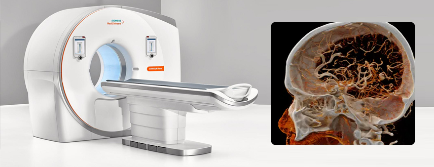
Our state-of-the-art CT systems utilize advanced imaging techniques to produce exceptionally detailed images of the brain, skull, spine, and surrounding structures in just minutes. High-resolution, low-dose CT technology helps physicians evaluate conditions such as stroke, traumatic brain injury, spinal fractures, hydrocephalus, brain hemorrhage, tumors, and degenerative spinal disorders. When appropriate, specialized contrast-enhanced imaging further improves visualization of blood vessels, inflammation, and other abnormalities, providing comprehensive diagnostic information while maintaining patient comfort and safety.
What are the benefits of Neurological CT?
- Rapid imaging during neurological emergencies
- Exceptional visualization of the brain, skull, and spine
- Advanced low-dose CT technology that minimizes radiation exposure
- Fast scan times with excellent patient comfort
- Accurate detection of bleeding, fractures, and other abnormalities
- High-resolution images that assist with diagnosis and treatment planning
- Expert interpretation by board-certified, fellowship-trained neuroradiologists
- Seamless collaboration with your referring physician for timely care
How does Neurological CT work?
During a neurological CT examination, you will lie comfortably on a motorized table that gently moves through the CT scanner while multiple X-ray images are captured from different angles. Powerful computer technology reconstructs these images into detailed cross-sectional views of the brain or spine, allowing radiologists to examine structures layer by layer. Most examinations are completed within minutes, are completely painless, and require little to no preparation, making neurological CT one of the fastest and most effective tools for diagnosing a wide range of neurological conditions.
First, the technologist ensures that you have no metal on your body, and reviews your medical history with you. You are then asked to lie on the scanning table, which is moved into the center of the machine, and asked to stay as still as possible to ensure the clearest images. Most exams last only a few minutes, depending on the body part.
The exam is non-invasive and the machine never touches your body. The technologist is available via intercom should the patient have any concerns during the exam.
You can be assured that only the minimal amount of radiation is used to ensure images of diagnostic quality.
24 hours prior to your exam, do not have any caffeinated or decaffeinated beverages or food including, energy drinks, coffee, tea, dark chocolate or diet pills.
For men, do not take Viagra or similar medications for 3 days prior to the exam.
Continue to take all your regular medications.
On the day of the exam, do not eat 1 hour prior.
Please leave all jewelry at home. Do not apply lotion to skin prior to exam.
Please be aware that sometimes it is necessary to take a medication called a betablocker to help lower your heart rate. If this occurs, you may be at the office for 1 hour or more.