Nuclear Medicine at Zwanger-Pesiri

Our nuclear medicine department combines the latest advances in technology with highly trained nuclear medicine physicians, radiologists, cardiologists, nurses, and nuclear technologists. We have a long-standing reputation for excellence in diagnostic nuclear imaging on Long Island, and offer this test in both Nassau and Suffolk counties.

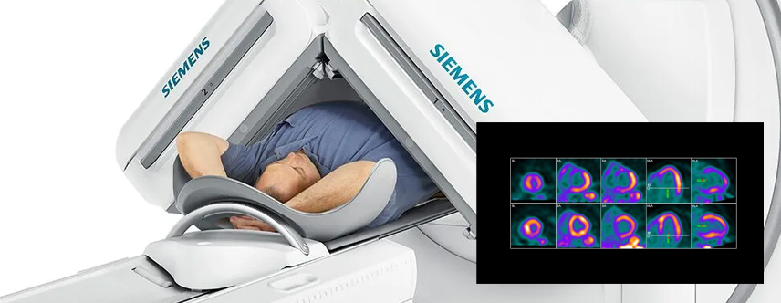
At ZP, we offer Single Photon Emission Computed Tomography (SPECT) imaging, which uses specialized cameras to visualize the internal organs at work, as well as the body's anatomy through three-dimensional images.

Nuclear medicine at Zwanger-Pesiri

What are the benefits of Nuclear Medicine

Pecise Insights for Better Health

Nuclear medicine imaging offers a unique window into the function of your organs and tissues, helping physicians detect, diagnose, and monitor a variety of medical conditions with remarkable precision. Unlike conventional imaging, which primarily shows structure, nuclear medicine provides detailed information about how your body is working at the cellular level. This allows for earlier detection and more personalized treatment planning.

Our advanced Nuclear Medicine provides patients with:
  • Early Detection: Identify diseases before symptoms appear.
  • Functional Insights: See how organs and tissues are working.
  • Targeted Treatment: Helps guide personalized therapy decisions.
  • Non-Invasive: Minimally invasive with low radiation exposure.
  • Comprehensive Monitoring: Track disease progression or treatment response accurately.

How does Nuclear Medicine work?

During a nuclear medicine scan, a small amount of radioactive material or radiopharmaceutical is introduced into your body either intravenously or orally and gets absorbed by the cells. A specially developed camera records images and measures the accumulation of the radiopharmaceutical in the patient’s body. Higher metabolic activity can correspond to areas of disease or “hot spots” in the study. For some studies, the activity of the radiopharmaceutical can indicate how well an organ is functioning.

Nuclear medicine can be used to detect and evaluate a number of disorders including tumors, irregular or inadequate blood flow, and inadequate functioning of organs like the thyroid, heart, lungs, gallbladder, liver, and kidneys.

Cardiac Stress Test

A cardiac stress test measures a heart's ability to respond to external stress in a controlled clinical environment. The stress response is induced by exercise or by drug stimulation. This exam is used to diagnose various symptoms, including chest pain, shortness of breath, or palpitations. It helps to identify abnormal heart rhythms, to see if enough blood flows to your heart, and help to find out if you have coronary heart disease.

Cardiac MUGA Scan

A MUGA scan (Multiple Gated Acquisition scan) is a noninvasive test used to evaluate cardiac function. The MUGA scan produces a moving image of the beating heart, and from this image, several important features can be determined about the health of the left and right ventricles (the heart’s major pumping chambers). A MUGA scan is particularly good at giving a reading of the overall pumping ability of the heart.

Nuclear Bone Scan

A nuclear bone scan uses a radioactive tracer and high-resolution camera to identify areas of new bone growth or areas in which bone has broken down. In general, bone scans are an excellent tool for identifying and evaluating damage to bones, assessing infections and trauma, and detecting cancer that has metastasized to the bones.

Frequently Asked Questions

For most exams, the radiopharmaceutical will be administered through an I.V. but some exams do require it to be taken orally. The radiotracer must circulate through your body for a certain amount of time, depending on the type of study. This can take anywhere from half an hour to several hours. The radiopharmaceutical is absorbed by both normal and abnormal tissue, according to their metabolic rate.

You will then be brought into the exam room and asked to lie down on the scanning table. A specialized nuclear medicine camera will slowly move over the area of your body being scanned. Be sure to remain as still as possible to ensure the best possible images. Depending on the specific study, your scan may take from 30 minutes up to two hours.

Once all of the images have been recorded, the nuclear camera will move away and the technologist will return to assist you off the table.

The specific test that you are having will determine the preparation required. Be sure to review the instructions given by the ZP representative, as well as your doctor, when scheduling your appointment. Some procedures require that you fast, while others require no special preparation.

Please call 631-444-5544, ext. 4886 to schedule and receive your preparations.

Increase your fluid intake for the next 24 hours to help flush the radiopharmaceutical out of your system. Depending on the type of exam, you may be required to stop breastfeeding or limit your contact with pregnant women, small children, and pets.