PET/CT at Zwanger-Pesiri

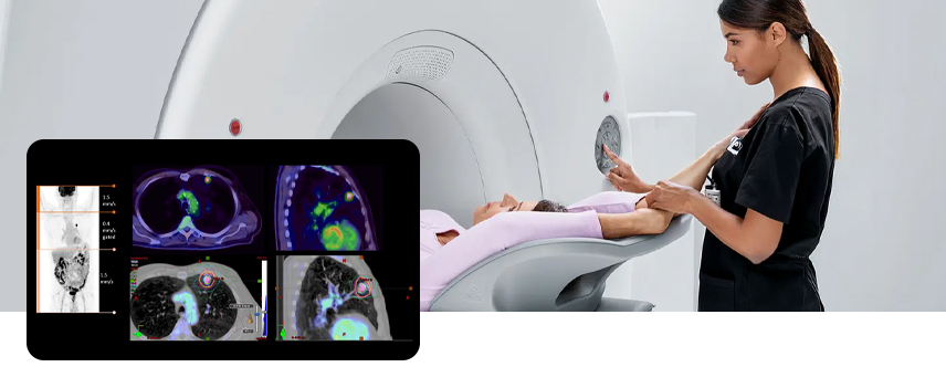
A PET/CT scan combines a PET scan and a CT scan to produce detailed images that help doctors more accurately detect and locate cancer, while increasing patient comfort. This combined imaging shows both the body’s structure and areas of abnormal metabolic activity, such as malignant tumor cells. By integrating the two scans into a single exam, PET/CT scans provide more accurate diagnoses than when PET and CT scans are performed separately.

PET/CT scan

How does PET/CT work?

The PET Portion

For the PET portion of the study, a small amount of radioactive material is injected into the body. The type of radioactive material depends on the exam. Once the injection is completed, the patient will wait about 60 minutes in a quiet area with limited movement while the body absorbs the material. More of the radiotracer material will accumulate in the cells with higher chemical activity, which generally corresponds to the areas of disease.

The CT Portion

For the CT portion, x-rays are taken from multiple angles as a patient is moved through the opening of the machine. An X-ray tube and high-resolution digital detector rotate very fast inside the machine’s opening to obtain pictures from all different angles and produce detailed images. The rotation of these parts is internal and cannot be detected by the patient. The images produced from a CT scan are significantly more detailed than a traditional X-ray.

PET/CT for Oncology

Physicians utilize PET/CT scans for diagnosing, staging, and evaluating treatments for their cancer patients. PET imaging provides the physician with information that can characterize a questionable abnormality as malignant or benign. CT provides detailed information about the location, size, and shape of various lesions. PET/CT captures images of changes in the body's metabolism which are caused by actively growing cancer cells. PET/CT reveals the size, shape, and exact location of any abnormal and cancerous growths.

PET/CT for Neurology

PET has been used for neurological imaging for many decades. It has grown in clinical importance, beyond its use in research, particularly due to patients with suspected dementia. Physicians can utilize PET imaging to visualize evidence of amyloid plaques when evaluating for Alzheimer's disease. PET/CT imaging can show precise areas of increased or decreased radiotracer uptake in the brain and doctors use this information to diagnose brain diseases, such as Alzheimer’s, dementia, and epilepsy.

PET/CT for Cardiology

PET/CT imaging of the heart enables the evaluation and measurement of multiple aspects of heart tissue function. Advances in PET/CT technology and specialized software have established PET/CT as an important tool in cardiology, supporting accurate diagnosis, detailed disease characterization, and effective treatment planning. It helps determine the severity of heart disease, measures blood flow and flow reserve, and distinguishes between viable and nonviable heart tissue.

PET/CT for Prostate Cancer

Prostate cancer is one of the most common cancers in men. ZP is now offering prostate-specific membrane antigen (PSMA) PET imaging, which significantly improves how prostate cancer is detected and treated.

A PSMA test uses a specialized tracer that is injected and attaches to PSMA proteins. Prostate cancer tumors over-express this protein, so the PET scan can detect the concentrated PSMA tracer.

This specialized test offers more precise detection of prostate cancer for better treatment planning and targeted care. Learn more.

Frequently Asked Questions

24 hours prior to the PET/CT scan

Refrain from any strenuous activity and do not eat or drink any caffeinated products (no coffee, tea, soda, chocolate, etc.), decaffeinated products, or juice. Eat a low carbohydrate diet (no bread, pasta, potatoes, rice, cereals, beans, or sweets). Chicken, fish, eggs, beef, cheese, bacon, and green vegetables are all okay to eat.
On the day of the PET/CT scan

6 hours before your exam:
  • 🚫  Do not eat any food.
  • 🚫  Do not drink anything.
  • 🚫  Do not smoke.
  • 🚫  Do not chew gum or tobacco.
On the morning of your exam:
  • 💧 You may drink small amounts of water.
  • 💊 You may take medications, only if they do NOT need to be taken with food.

Remove all jewelry and any other metallic objects such as metal zippers, body piercings, and removable dental work. Wearing comfortable clothing with no metal may prevent you from having to change into a gown.

Refrain from any strenuous activity and do not eat or drink any caffeinated products (no coffee, tea, soda, chocolate, etc.), decaffeinated products, or juice. Eat a low carbohydrate diet (no bread, pasta, potatoes, rice, cereals, beans, or sweets). Chicken, fish, eggs, beef, cheese, bacon, and green vegetables are all okay to eat.

After reviewing your medical history, the nurse or technologist will place an intravenous needle into a vein in your arm or hand. Your blood sugar level will then be checked and the radioactive tracer will be administered through the I.V.

You will then be brought into the PET/CT room and asked to lie down on the scanning table. The scanning table you are lying on will be moved into the circular opening in the middle of the machine and the test will begin. The exam is non-invasive and the machine never touches you. Be sure to remain as still as possible to ensure the best possible images. The entire scanning portion of the exam takes about 20 to 30 minutes and your technologist will be able to talk to you from outside the room through an intercom.

Once all of the images have been recorded, the scanning table will move out of the PET/CT machine and the technologist will return to assist you off the table.

If you have impaired kidney function, are diabetic or are 60 years of age or older, we will perform an i-STAT creatinine level at the time of your exam to assess your kidney function. It is important to inform us if you are taking the medication hydroxyurea when making your appointment. Keep hydrated before and after your exam.