Diagnosing Neurodegenerative Diseases with NeuroQuant®
Zwanger-Pesiri Radiology now offers NeuroQuant® AI software, further advancing the capabilities of its expert Neuroradiology department with some of the latest MRI technology available across Long Island. This cutting-edge software uses artificial intelligence to analyze brain MRI scans with precise volumetric measurements that can assist physicians in evaluating structural brain changes associated with a range of neurodegenerative diseases, including Alzheimer’s disease, dementia, and other memory-related conditions.
By staying at the forefront of innovation, ZP continues to provide patients and referring physicians with advanced Wide Bore MRI technology to support earlier detection, more accurate assessments, and confident treatment planning.
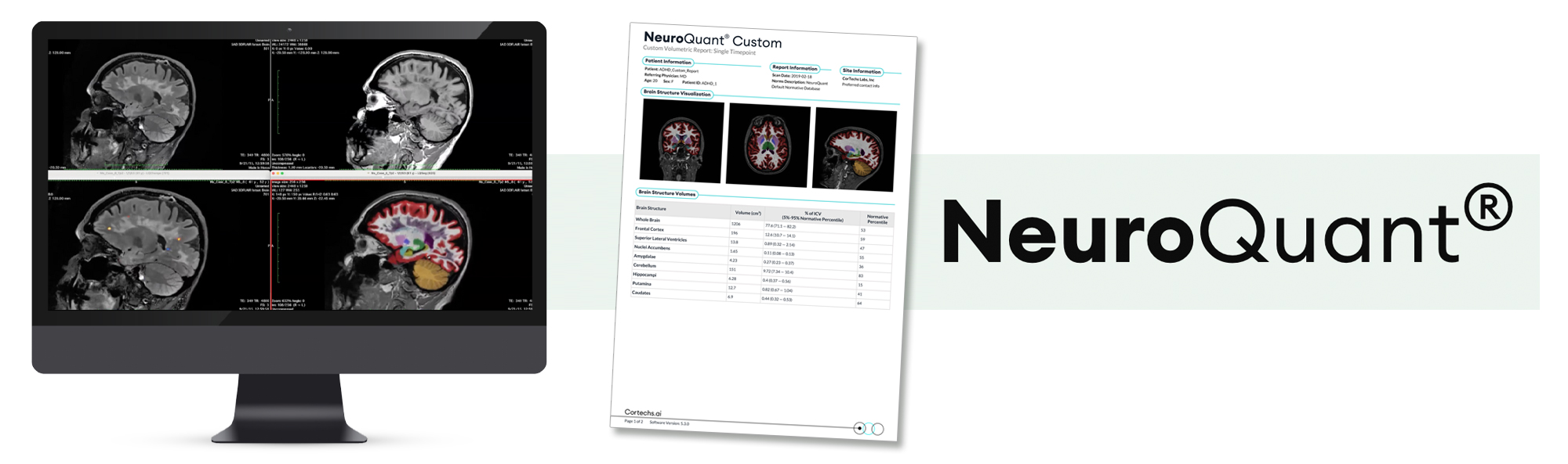
Better Accuracy with NeuroQuant®
NeuroQuant® provides clinicians with accurate segmentation, quantification, and volume measurements of the brain. MRI acquisitions are sampled to detect pathological changes and atrophy, as well as to support lesion surveillance.
- Uses conventional MRI images to process and quantify brain measurements
- Comprehensive reporting includes multi-structured brain segmentation and lesion counts
- Monitor lesion counts and maximum diameters over time
- Added clinical value to MRI reports
- Improve patient monitoring and treatment
Identifying neurodegeneration in its earliest stages
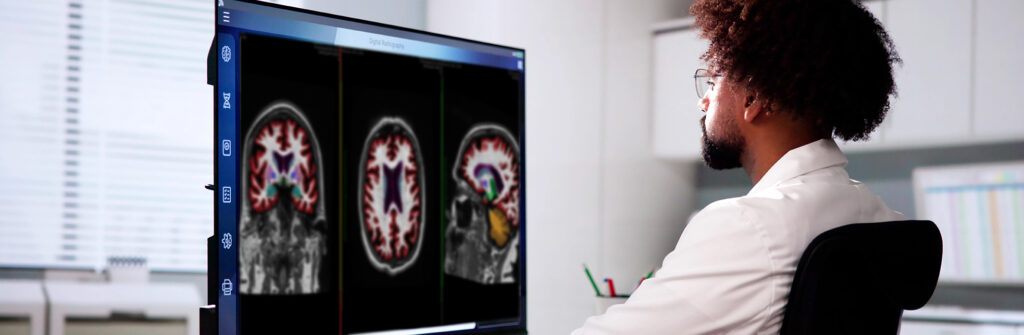
ZP is assisting physicians to provide the best treatment and lifestyle planning for patients by supporting their clinical impression with objective, volumetric data and color-coded brain structure analysis. With NeuroQuant®, we can now increase diagnostic accuracy and reduce variability between diagnostic impressions.
Frequently Asked Questions
If you are receiving IV CONTRAST for your exam, have nothing to eat 1 hour prior to your exam time. You may drink clear liquids (example: water, ginger ale, apple juice).
Because the MRI system uses a very powerful magnet, there are many items that cannot be brought into the exam room. Although a private locker will be provided, it is best to leave certain items at home or with someone during your MRI exam, including:
- Cell phones, pagers, iPads, or any other electronic device
- Credit cards, driver’s license, and any other card with a magnetic strip
- Loose change
- Bobby pins, safety pins or any other item with metallic pieces
- Any loose metallic items on clothing or body
- Glasses, hearing aids and items such as TENS Units will not be able to go into the exam room, but can be worn until just before entering the MRI room.
- Wear comfortable loose-fitting clothes, such as a sweatsuit. Be sure there are no metal zippers, snaps or buckles. Do not wear earrings, hairpins, or jewelry. Do not apply eye shadow or mascara.
This exam may not be performed if you have a cardiac pacemaker, cerebral aneurysm clips or a metallic hearing implant. If you are a sheet metal worker or have ever had metal fragments in your eye(s), an orbit x-ray will be taken prior to your MRI exam.
After checking in, you will be escorted to a changing room. You will then meet the registered MRI technologist who will be performing your exam. The technologist will review the screening questionnaire and the procedure with you, answering any questions you may have.
When you are ready, you will be asked to lie down on the cushioned MRI table. Many MRI exams require the use of a coil, which is placed on or around the body part being scanned. The coil is a lightweight device that acts as an antenna to receive the radio frequency signal coming out of your body. That data is then transmitted to a computer, which will then generate the images. If it has been determined that your study requires the use of a contrast material (or dye), the technologist or nurse will perform the injection and administer the iodine-free contrast. Please let the staff know if you have any allergies.
The table will slowly move into position within the large doughnut-shaped magnet. We provide you with noise-canceling headphones and your choice of music, as you will hear a series of loud thumping noises as the scan is performed. With our modern MRI systems, most exams are over in 3 songs! We ask that you keep very still to help ensure that the images come out clear. Throughout the entire exam, you will be able to communicate with the technologist through an intercom.
An MR Angiography exam can take anywhere from 10 to 30 minutes.