Orthopedic CT at Zwanger-Pesiri
Zwanger-Pesiri Radiology's Orthopedic Imaging Department excels in utilizing computed tomography (CT) for orthopedic issues, showcasing expertise and precision in diagnosing complex bone and joint conditions. Their specialized approach involves the latest CT technology, allowing for detailed, cross-sectional images that are crucial for accurate diagnosis and treatment planning.
The department's skilled radiologists are adept at interpreting CT scans of bones and joints, identifying fractures, evaluating bone density, and detecting abnormalities with exceptional accuracy. Their proficiency in CT imaging ensures that patients receive the highest standard of care, leading to more effective treatment outcomes and improved overall orthopedic health.
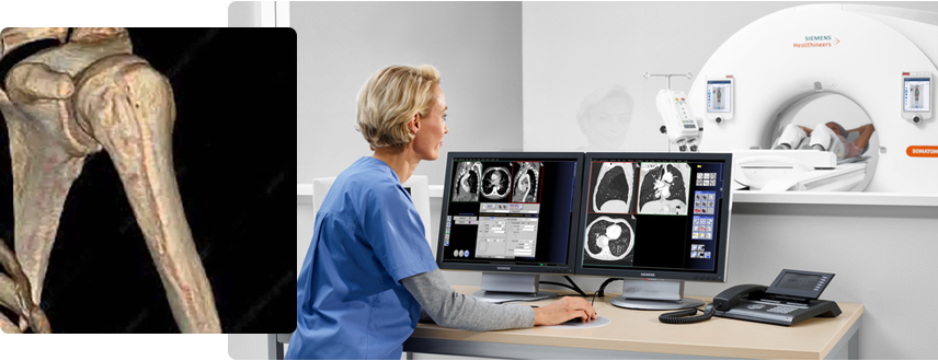
What are the benefits Orthopedic CT imaging?
Computed tomography (CT) is a valuable imaging modality in orthopedics, offering several benefits over traditional X-rays and other imaging techniques. One of the key advantages of CT in orthopedic imaging is its ability to provide detailed, cross-sectional images of bones, joints, and surrounding soft tissues. This high level of detail allows for more accurate diagnosis and treatment planning, particularly in complex cases or when traditional X-rays are inconclusive.
CT is also particularly useful for imaging complex anatomical structures, such as the spine and joints, where precise visualization is critical. It can help orthopedic surgeons evaluate fractures, assess bone density, and detect abnormalities such as tumors or infections. Additionally, CT can be used to guide orthopedic procedures, such as bone biopsies, joint injections, or the placement of screws or implants.
Another benefit of CT is its speed and efficiency. While magnetic resonance imaging (MRI) is often considered the gold standard for soft tissue imaging, CT scans can be performed more quickly, making them ideal for patients who cannot tolerate longer imaging sessions or who require urgent evaluation.
When used judiciously and in combination with other imaging modalities when necessary, CT can be an invaluable tool in the field of orthopedics, helping to improve patient outcomes and quality of care.
Who can benefit from Orthopedic CT imaging?
- Individuals with complex fractures or injuries requiring detailed imaging
- Patients with suspected bone tumors or infections
- Those needing pre-surgical planning for joint replacement or corrective surgery
- Athletes with sports-related injuries requiring precise diagnosis
- Patients with chronic orthopedic conditions requiring ongoing monitoring
- Individuals with spinal abnormalities or degenerative conditions
- Those needing guidance for orthopedic procedures such as biopsies or joint injections
First, the technologist ensures that you have no metal on your body, and reviews your medical history with you. You are then asked to lie on the scanning table, which is moved into the center of the machine, and asked to stay as still as possible to ensure the clearest images. Most exams last only a few minutes, depending on the body part.
The exam is non-invasive and the machine never touches your body. The technologist is available via intercom should the patient have any concerns during the exam.
You can be assured that only the minimal amount of radiation is used to ensure images of diagnostic quality.
24 hours prior to your exam, do not have any caffeinated or decaffeinated beverages or food including, energy drinks, coffee, tea, dark chocolate or diet pills.
For men, do not take Viagra or similar medications for 3 days prior to the exam.
Continue to take all your regular medications.
On the day of the exam, do not eat 1 hour prior.
Please leave all jewelry at home. Do not apply lotion to skin prior to exam.
Please be aware that sometimes it is necessary to take a medication called a betablocker to help lower your heart rate. If this occurs, you may be at the office for 1 hour or more.