DEXA at at Zwanger-Pesiri
A DEXA bone densitometry exam is the most accurate and most commonly used method for measuring bone loss. It is the best way for doctors to diagnose conditions like osteoporosis or osteopenia, as well as assess a patient’s risk of suffering a bone fracture.
DEXA exams are primarily recommended if you have x-ray evidence of vertebral fracture, a family history of osteporosis or hip fracture, or have experienced a fracture after only mild trauma.
If you have hyperthyroidism, hyperparathyroidism, type 1 diabetes, use medications that are known to cause bone loss, or are a post-menopausal woman not taking estrogen, your doctor may recommend a DEXA exam.
The benefits of DEXA Bone Densitometry?
- Accurately measures bone density to help diagnose osteoporosis and assess fracture risk.
- Quick, painless, and noninvasive, typically completed in just a few minutes.
- Uses very low-dose X-rays, making it one of the safest imaging tests available.
- Monitors bone health over time, allowing providers to evaluate the effectiveness of treatments.
- Identifies early bone loss, enabling proactive steps to protect long-term skeletal health.
- Provides precise, reliable results, essential for guiding personalized care plans.
What should I expect during a DEXA scan?
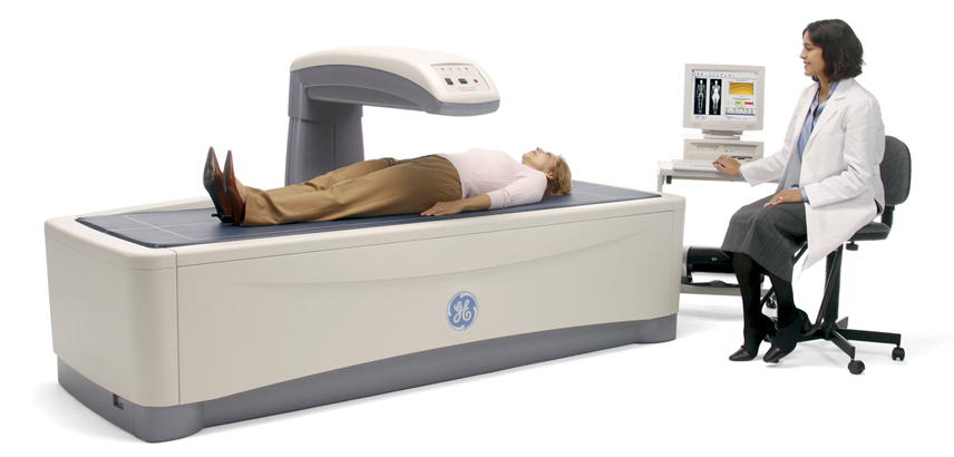
During a DEXA scan patients lie on their backs and the machine passes over them. It is a non-invasive, painless procedure that measures your bone density. The machine uses very low dose x-rays with differing energy levels that get directed at the bones being scanned. The images produced allow the radiologist to determine your bone mineral density. Fewer x-rays pass through denser bone. This information tells doctors the average density of the bone. A low score indicates that the bone is more prone to fracture and some material of the bone has been lost.

Bone Health
Know your risk of a fracture before its too late
Easy exam
Easy and convenient procedure with minimal radiation
Expert Care
Women's Imaging Radiologists for expert diagnosesLearn about the different types of DEXA exams
Vertebral Fracture Assessment
Vertebral Fracture Assessment (VFA) examines the bones of the spine to determine whether any vertebrae have an abnormal shape or compression fractures. This exam can be performed during the same visit as a DEXA scan and is especially helpful for individuals at high risk for vertebral fractures. If you have noticed a loss of height over time, developed a stooped or hunched posture, experienced a fracture as an adult, or have unexplained back pain, you may have a vertebral (spinal) fracture.
Prior vertebral fractures are a stronger predictor of future fractures than low bone mineral density alone. Patients with vertebral fractures face a significantly increased risk of additional fractures. Nearly two-thirds of vertebral fractures occur without noticeable back pain, making this test especially valuable.
FRAX® Tool
Vertebral Fracture Assessment
The FRAX tool was developed by the World Health Organization to evaluate the risk of fractures for patients with low bone mass. The FRAX® models have been developed from studying population-based cohorts from Europe, North America, Asia and Australia. FRAX® can help to identify people who have a greater chance of breaking a bone as well as people who might benefit from taking osteoporosis medicine. The tool looks at a person’s age, family history, bone density and other factors to estimate the patient’s chance of fracturing a hip or another major bone within the next ten years. It can be used to guide treatment decisions in postmenopausal women, men over the age of 50, and people with low bone density.
You must remove all jewelry and any other metallic objects such as hearing aids, jeans with metal zippers, and body piercings.