Breast Ultrasound at Zwanger-Pesiri
Breast ultrasound imaging uses sound waves to produce pictures of the internal structures of the breast. This procedure is primarily done to help diagnose breast lumps or other abnormalities your doctor may have found during a physical exam, mammogram, or breast MRI. Breast ultrasounds are safe, non-invasive, and do not use any radiation. This test is often administered when a change has been seen on a mammogram or when a change is felt but does not show up on a mammogram.
Breast ultrasound is not used as a primary screening tool for breast cancer, as it may miss some early signs of the disease. Certain early changes in breast tissue may not be visible on ultrasound, so additional imaging methods or diagnostic procedures are often needed to make an accurate diagnosis.
Why might I need a Breast Ultrasound?
- You have particularly dense breast tissue.
- A mammogram may not be able to see through this tissue.
- You are pregnant. Ultrasound does not use any radiation, making it safer for the fetus.
- You are younger than age 25.
- Your healthcare provider wants to look at nearby lymph nodes.
- Your healthcare provider needs to use ultrasound to guide a needle during a biopsy.
How does Breast Ultrasound work?
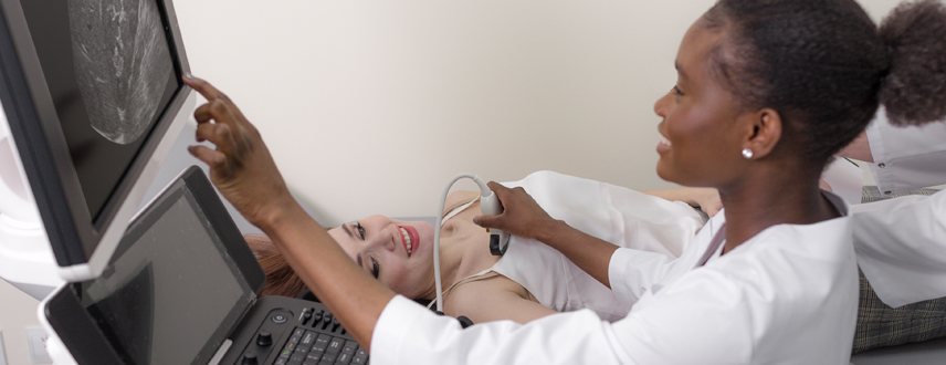
Breast ultrasound works by using high-frequency sound waves to create detailed images of the breast tissue. During the exam, a technologist applies a warm gel to the skin to help transmit the sound waves, then gently moves a handheld device called a transducer over the breast. The sound waves bounce off internal structures and return to the transducer, where they are converted into real-time images displayed on a monitor. This allows radiologists to closely evaluate areas of concern—such as lumps, cysts, or dense tissue—and distinguish between fluid-filled and solid findings. It is a safe, painless, and radiation-free tool that provides valuable diagnostic insight.
Your technologist will move a wand-like device, called a transducer, over your skin to take images of your breasts. The transducer sends out waves that bounce off your breast tissue. The sound waves are too high-pitched for you to hear, but the transducer can pick up the bounced sound waves, which are then made into pictures of the inside of your breasts.
Depending on your procedure, your technologist may also use a Doppler probe, which allows the radiologist to assess blood flow using sound waves emitted by the transducer. This helps to determine how fast blood is moving through a vessel and the direction of flow. Little or no detectable sound may indicate reduced or blocked blood flow.
Before beginning the procedure, your healthcare provider will explain the process and answer any questions you have. You do not need to stop eating or drinking before the test. Do not apply any lotion, powder, or other substances on your breasts the day of the test. You will be given a gown to change into, so please wear clothes that can easily be removed.