MRI at ZP
Long Island's Leader in MRI
Zwanger-Pesiri Radiology has been leading the way in high-end MRI (Magnetic Resonance Imaging) technology for decades, with more experience than any hospital or radiology center in NY. We offer the latest in 3T and 1.5T Wide Bore MRI, as well as our Open MRI Solutions at our Massapequa and Coram offices. IV sedation is available for those with extreme claustrophobia or anxiety.
Our MRI technology is updated every two to three years, guaranteeing patients will never be scanned on outdated equipment, while always receiving the sharpest images in the shortest time. We provide a variety of technology as every patient is different and deserves the most comfortable MRI experience possible.
ZP invests in the latest innovations from Siemens Healthineers to offer a unique combination of comfort and image quality for your MRI exam.

3T & 1.5T Wide Bore MRI
Open Sided MRI
Open Wide MRI
What are the benefits of MRI?
- Produces highly detailed images of soft tissues, organs, and structures.
- Uses no ionizing radiation, making it a safe imaging option.
- Excellent for evaluating the brain, spine, joints, and abdominal organs.
- Helps in early detection of tumors, injuries, inflammation, and other abnormalities.
- Provides multiple imaging sequences for a comprehensive view of the body.
- Useful for diagnosing complex conditions that other modalities may miss.
- Noninvasive and painless.
- Offers advanced techniques such as functional MRI and MR angiography for specialized evaluations.

How does MRI work?
MRI is a non-invasive imaging technology that produces three-dimensional detailed anatomical images without the use of damaging radiation. It is often used for disease detection, diagnosis, and treatment monitoring. It is based on sophisticated technology that excites and detects the change in the direction or the rotational axis of protons in the water that makes up living tissues.
The MRI scanner takes multiple very thin two-dimensional pictures, which the computer assembles into three-dimensional pictures. This allows the doctor to look layer by layer at the area being scanned, which provides greater detail in the diagnostic process.

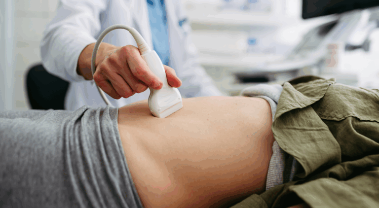
What can I expect during an MRI exam?
You will be brought into the MRI room and asked to lie down on the scanning table. The area of your body being scanned will be comfortably positioned in or near a special surface coil.
You will be moved into the center of the magnet and the test will begin. The machine never touches you and the exam is completely non-invasive. You will be asked to remain still to ensure the best possible images. The technologist will be right outside the room while keeping you aware of your progress through an intercom. Once the images have been recorded, you will be taken out of the MRI machine and the technologist will return to assist you off the table.
Please note that MRI machines are loud. All of our MRI suites were carefully constructed using extensive sound proofing acoustical material. This helps reduce the noise throughout the office, as well as lowering the noise level for the MRI patients up to 15 decibels! In addition, every MRI patient is offered noise cancelling headphones so they can listen to music during their scan. Due to the advanced machinery and fast scan times, most patients are done by the third song!

At ZP, we use the most advanced technology, and have the fastest scans
MRI uses zero radiation
During the exam, you get headphones with your choice of music
We offer a wide range of machines for MRI
New Metal Reduction Free.Max MRI
This groundbreaking technology offers major improvements for imaging patients with metal implants, as implanted devices cause artifacts when scanning with MRI. Also, this new MRI offers the world’s first 80cm bore, for claustrophobic and anxious patients. Learn more about Free.Max MRI from Siemens.
Elastografía Hepática
Ahora ofrecemos exámenes de ultrasonido con Elastografía Hepática El avanzado departamento de ultrasonido de…

Mamografía 3D
Mamografía 3D en ZP Su mamografía de detección anual es uno de los pasos más…

Food Donation to Pronto of Long Island
Zwanger-Pesiri Radiology, Long Island’s leading provider of diagnostic imaging services, is proud to announce the…
ZP Hauppauge Grand Opening
Zwanger-Pesiri Radiology is proud to announce the grand opening of our newest state-of-the-art location in…

Best of Long Island 2025
Zwanger-Pesiri Radiology is deeply honored and grateful to the Long Island community for voting us…
How to Prepare for MRI
MRI uses a very powerful magnet. It is extremely important that you do not bring any metal into the area. Doing so can create a dangerous scenario for you and staff members. You must remove all jewelry and any other metallic objects such as hearing aids, jeans with metal zippers, body piercings, and removable dental work. Wearing a sweatsuit with no metal may prevent you from having to change into a gown.
Why Choose Zwanger-Pesiri?
Zwanger-Pesiri Radiology brings world-class expertise to the Long Island community. Our subspecialty-trained radiologists are Board Certified by the American Board of Radiology with fellowship training in a variety of specialties. They are highly-skilled, highly-knowledgeable, and make patient care a priority. To learn more, contact us today.