CT at ZP
Zwanger-Pesiri is dedicated to providing Long Island with the latest CT (Computed Tomography) technology, offering 256, 128 and 64-slice systems. We are able to greatly reduce the radiation dose to patients while still maintaining the highest resolution. With our 256-slice CT-Flash scanners in Plainview and Stony Brook, patients are exposed to 75% to 90% less radiation than from most other CT scanners.
ZP participates in Image Wisely™, a campaign that encourages smart medical imaging. We pledge to eliminate unnecessary scans and lower radiation doses by using state-of-the-art equipment.
Each CT patient at ZP receives a card indicating the actual radiation dose that they were exposed to. We encourage patients to hold onto this card as part of their personal medical record.
What is a CT?
CT stands for Computed Tomography. It is a safe and painless test that uses x-rays taken from different angles to produce detailed images of bones, soft tissue, organs and blood vessels. The images produced from a CT scan are significantly more detailed than a traditional x-ray. CT has revolutionized areas such as cardiology, neurology, orthopedics and oncology.
CT may be used to evaluate many conditions including broken bones, cancer, blood clots, internal bleeding and signs of heart disease.
How does CT work?
During a CT scan, you are placed on a table which slides into a large doughnut-shaped opening. A powerful x-ray tube and high resolution digital detector rotate very fast inside the doughnut to obtain pictures from all different angles.
The CT scanner takes many very thin 2-dimensional pictures, which the computer can assemble into 3-dimensional pictures. This allows the doctor to look layer by layer at the area being scanned and provides greater detail to aid in the diagnostic process.
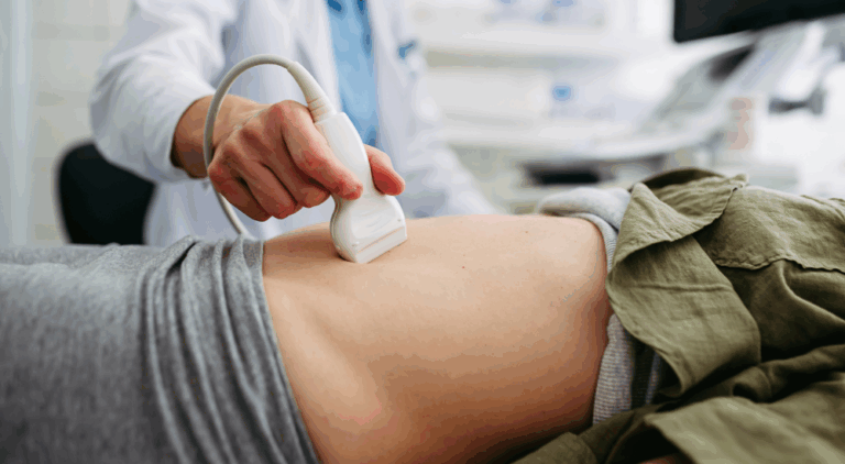
Many CT scans require the use of a contrast dye. The contrast may be a drink that you take prior to the scan or may be administered during the scan through an I.V. The contrast highlights certain parts of your body and helps to provide the sharpest images available.
How do I prepare for a CT scan?
When scheduling your appointment (online or by phone), provide us with your email address and we will send you personalized forms with your information already filled in. You will only have to update or add any missing information. If your email address is not provided, you can still save time by downloading the CT forms and completing them prior to arriving at the office.
You must remove all jewelry and any other metallic objects such as hearing aids, jeans with metal zippers, body piercings and removable dental work. Wearing a sweatsuit with no metal may prevent you from having to change into a gown.
Additional prep for CT scan with oral or I.V. contrast
Have nothing to eat 1 hour prior to your exam time. You may drink clear liquids (example: water, ginger ale, apple juice). Keep hydrated before and after your exam.
If you have impaired kidney function, are diabetic or are 70 years of age or older, we will perform an i-STAT creatinine level at the time of your exam to assess your kidney function. It is important to inform us if you are taking the medication hydroxyurea when making your appointment.
If you are receiving oral contrast, please pick up the contrast kit the day before your exam. If you are receiving Omnipaque oral contrast, refer to the Omnipaque oral contrast section below. If you are receiving Redi-CAT oral contrast, please ask your Zwanger-Pesiri representative for those specific instructions.
Omipaque oral contrast prep for CT scan
Do not take if you have an iodine allergy.
Begin drinking the Omnipaque oral prep 1 hour before your exam.
To prepare the contrast drink:
- Pour the entire contents of the Ominpaque bottle into the
32 oz. cup that was given to you. - Fill the cup with water up to approximately 1/2 inch from the top
of the cup (approximately 30 oz). - Stir well and drink.
- Discard the cup, contrast bottle and straw after use.
Bring with you to the appointment:
- Prescription from your doctor.
- Current insurance card.
- Authorization number from your insurance carrier.
- Any forms you completed at home.
- Credit card or cash for your insurance co-pay.
- Any studies of the same body area that you have from another facility. We like to compare the new CT study with any previous studies to assist in the diagnostic process.
- Picture identification.
Plan to arrive 15 minutes before your scheduled appointment time.
What happens during the test?
All metal must be removed before your scan including jewelry, dentures, eyeglasses, belt buckles and any clothing with metal zippers or buttons. Metal interferes with the quality and accuracy of the images captured during the CT. You may be asked to change into a gown.
The CT technologist will confirm that you are free of all metal and review your medical history with you.
You will then be brought into the CT room and asked to lie down on the scanning table. The scanning table you are lying on will be moved into the center of the opening and the test will begin. The machine never touches you. Be sure to remain as still as possible to ensure the best possible images. Although the CT technologist cannot stay in the room with you during the scan, he or she will be able to talk to you from outside the room through an intercom.
Once all of the images have been recorded, the scanning table will move out of the CT machine and the technologist will return to assist you off the table.
The CT scan can take from 2 to 10 minutes, depending on the area of the body being scanned.
For CT with I.V. contrast
After reviewing your medical history, the nurse or technologist will place the intravenous needle/catheter into a vein in your arm or hand. Then at a specific time in the exam, contrast will be injected into your I.V. It is normal to feel a warm sensation throughout your body and a metallic taste in your mouth.
What will I do when I arrive?
Present your prescription, insurance card and completed forms at the front desk. If any additional forms are required, they will be given to you at this time.
Be sure to inform the receptionist and technologist if you:
- Have allergies, specifically to iodine.
- Are pregnant or think you may be pregnant.
- Have any compromised kidney function or a history of kidney disease.
- Are currently taking any medications.
- Have any metal or a medical device in your body.
- Have diabetes.
- Have asthma.
- Have any studies of the same body area from another facility. We like to compare the new CT study with any previous studies to assist in the diagnostic process.
When can I expect the results?
Before leaving the office, you will receive a CD with the images from your CT exam.
One of our board certified radiologists specializing in CT interprets your images, compares them to any previous studies and dictates a report which is transcribed, proofread and signed.
The report is then faxed and mailed to your referring doctor within one or two days.
Your doctor will read the report and review the findings with you.
All of your signed reports and images are available to your referring doctor on our physician’s web portal.
256-Slice CT-Flash
The CT-Flash scanner sets new standards regarding speed and radiation dose reduction. This system features the latest in dual-source CT technology, utilizing two x-ray sources that rotate simultaneously around your body. We are able to scan the heart in only 250 milliseconds (a quarter of a heartbeat) and the full body in only seconds. Scanning patients in a flash allows us to greatly reduce the radiation dose while still maintaining superb image quality.

Compared to most other scanners being used today, the CT-Flash exposes patients to 75% to 90% less radiation. Compared to a nuclear stress test, patients are exposed to 95% less radiation. Not only through its unmatched speed are we able to emit such low doses, but through specialized adaptive dose shields that help to block any unnecessary radiation. Furthermore, the CT-Flash's unique ability to turn off the x-ray beam while pointed at radiation sensitive organs, like the breast and thyroid, lowers the dose even more.
CT Angiography
CT Angiography (CTA) is a radiological test that is used to create detailed images of the blood vessels in your body, including those in your heart, lungs, kidneys, legs and arms.
A CTA is commonly used to examine pulmonary arteries in the lungs, detect clots in the veins, visualize blood flow in the renal arteries and identify aneurysms inside the brain. During a coronary CTA, we check for narrowed arteries in your heart (coronary heart disease) that could put you at risk for a heart attack.

This minimally invasive procedure involves injecting contrast dye into a small vein in your arm or hand through an I.V. During a coronary CTA, you may be given betablockers or nitroglycerin to control your heart rate. The CT scanner then uses a series of x-rays to obtain many cross-sectional images which are reconstructed by a computer into very detailed 3-dimensional pictures. A CTA is an alternative to a catheter angiogram, a much more invasive procedure that uses a large catheter to inject the contrast material, usually through the groin.
You can be assured that at Zwanger-Pesiri, we utilize the latest in CT technology to produce images of extraordinarily fine detail using the minimum amount of radiation.
Dental CT
Previously, radiographic images that were used to place implants were two dimensional. These two dimensional images provided a reasonable estimate of bone height, but often bone width was not determined until the bone was exposed during surgery. This often led to surprises for the implant surgeon, such as inadequate bone structure.

Today, we offer the latest in dental CT scanning, which provides detailed 3D images of the structures of the jaw. These 3D visualizations, coupled with specialized software, allow implants to be placed with a level of precision that was unattainable years ago. Dental implants can now be accomplished in a much more conservative way, which involves greatly reduced discomfort, less treatment time and a more accurate final outcome.


